Evaluation of anterior-posterior length of eyeball

Evaluation of anterior-posterior length of eyeball

Ophthalmologists are able to assess the anterior-posterior length of the eyeball using advanced diagnostic techniques.

IOL Master

It is an advanced device that can be used to measure the anterior-posterior length of the eye and the curvature of the eye surface in one minute without contact with the eye. Therefore, using this device does not have the risks associated with contact with ocular devices and does not require anesthesia to use it.

Examination of the retina and blood vessels

  1. angiography:

    To perform this test, a contrast agent, such as fluorescein or the like, is injected into one of the patient’s blood vessels. The FUNDUS tool is used to photograph the back of the eye.

    This test is used to check for retinal and choroid blood flow. Fluorescein is commonly used to examine retinal arteries and ICG is used to examine choroidal arteries and deeper areas. Fluorescein angiography is mostly used to evaluate diabetic retinopathy, obstructive vascular diseases such as occlusion of arteries or retinal veins, and to evaluate wet macular degeneration.

    OCT:

    This new technique is known as Optic Coherence Tomography. Using OCT, the ophthalmologist obtains high-precision tomographic sections of the retinal layers and other valuable information. It is therefore used to diagnose and follow many retinal diseases such as macular perforation, macular edema, macular degeneration, diabetic retinopathy and glaucoma.

    Since this technique uses a light source, there is no need for eye contact and it is done in a few seconds.

Corneal examination

The cornea is the transparent part in front of the eyeball that separates the inside of the eye from the outside like glass. The cornea has two important functions: one is to direct light rays into the eye and focus them on the retina, and the second is to protect the structures inside the eyeball.

In order for light to enter the eye and reach the retina, it must first pass through the cornea. Therefore, the clarity of the cornea is very important in vision, and any problem can disrupt the function of this very important organ of the visual system. Ophthalmologists use various diagnostic methods to identify these problems and guide the patient through the treatment process.

Today, with advances in ophthalmology, highly advanced tools, equipment, and techniques have been developed to evaluate and evaluate the structure and function of the cornea. These tools include:
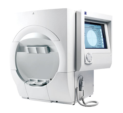
Topography

Using the topographic method, light rings are sent to the surface of the cornea; By receiving the reflection of these rings and analyzing the received information and examining the obtained topographic map, any corneal disorders such as keratoconus, corneal scar and corneal curvature can be identified. This procedure is a major procedure for people who want to undergo refractive surgery such as LASIK, LASEK and PRK. It may also be used in postoperative follow-up in some people.
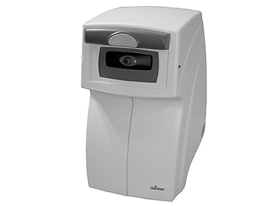
Pachymetry

Metric purity is a method in which the thickness of the cornea is measured using ultrasound waves. Metric cleansing is a basic procedure before refractive surgery because in such surgeries, part of the corneal tissue is removed.

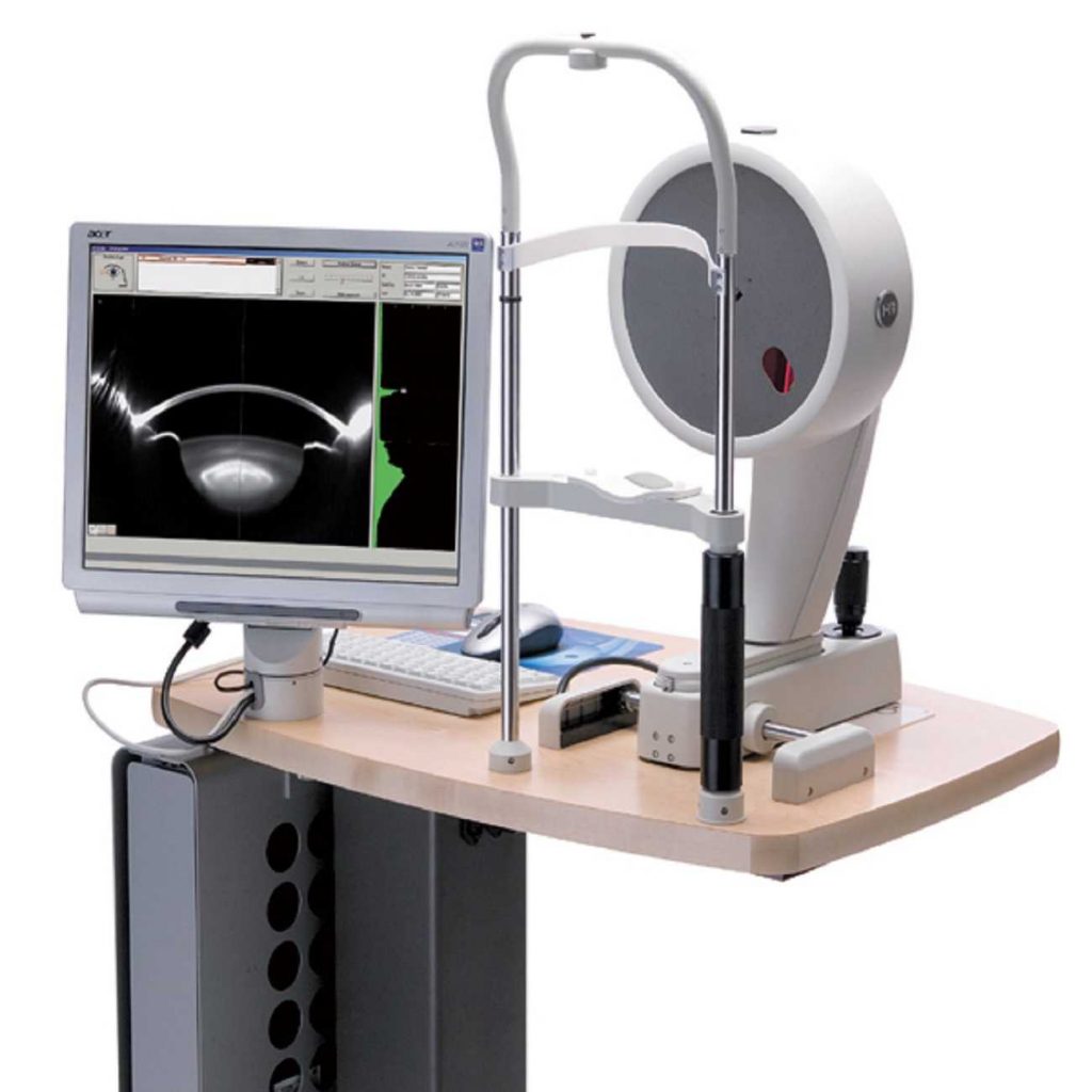
Pentacam and Orbscan

By Pentacom and Arb scanning device, the topography of the cornea, ie the general structure of the cornea, including its surface strength, thickness and shape of its front and back parts, is determined. This is one of the techniques that can evaluate the posterior part of the cornea.

Check the visual field

The field of vision is a range of space that can be seen by a person with a fixed gaze in one direction.

The main use of this test is to diagnose and monitor glaucoma. It also helps diagnose a variety of eye diseases and visual pathways in the brain.

If your doctor recommends this test, it means that your eye nerve is suspected; This suspicion can be due to ocular or neurological problems, the most important and most common of which are increased intraocular pressure (such as glaucoma), damage to the optic nerve pathway, increased cerebrospinal fluid pressure, and inflammation of the optic nerve head.

The visual field test can be performed in a clinical examination by asking the patient to stare at a point and keep the eye steady, while moving an object like a pen in different parts of the visual field.

In this examination, we calculate the angle at which a person is able to see and perceive an object. When computer devices are used for testing, the diagnosis will be accurate and reliable. A perimeter or hemisphere device in which points of light flash on a white screen is measured.